Author: Chris Janse
<< previous chapter | next chapter >>
See PDF-file for a number of light microscope images of different P. berghei life cycle stages
A description of the (light microscopy) morphology, including pictures of different life-cycle stages can also be found in Landau, I. And Boulard, Y. (1978) Life cycles and Morphology. In: Rodent Malaria (R. Killick-Kendrick and W. Peters, eds.) pp. 53-84. Academic Press, London.
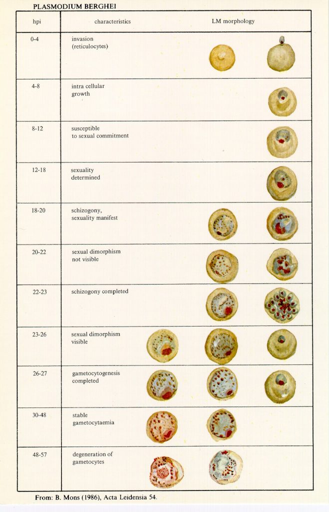
Figure: Light microscope morphology of blood-stages of P. berghei (Giemsa-stained).
Blood-stages shown are from synchronized blood-stage infections of the ANKA strain of P. berghei at several time points after invasion (hours post invasion – hpi) of merozoites into erythrocytes (from: Mons, B (1986) Acta Leiden. 54, 1-124).
Transmission electron microscope images of various blood-stages of P. berghei
The emphasis is on the development of gametocytes. The blood stages of P. berghei ANKA were obtained from synchronized infections at several time points after invasion of erythrocytes (hours post invasion – hpi). Magnification 16.000-17.000x. (from Mons, B (1996) Acta Leiden. 54, 1-124).
See PDF-file for larger images
See PDF-file for some transmission electron microscope images of the mature ookinete stage of P. berghei


